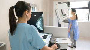
When it comes to technology, Artificial Intelligence (AI) is the newest frontier where industries are starting to invest their time and energy.
Healthcare organizations, including Rochester Regional Health, are beginning to incorporate AI into different areas of care, including breast cancer screenings.
Shruthi Ram, MD, is the Section Chief of the Division of Breast Imaging with Rochester Regional Health and describes how AI is helping radiologists and imaging providers serve their patients even better.
AI trains a machine to perform certain tasks by feeding it massive amounts of data. For breast cancer imaging, using AI allows the machine to analyze images and detect cancers at a much faster pace than a human would be able to learn. Over the course of a radiologist’s career, they might review 30,000-40,000 mammograms. AI can review millions of images and compare them against one another in a fraction of the time it would take a human being.
Rochester Regional Health is partnering with Volpara Analytics at breast imaging centers to use AI-integrated software. The software offers a way to better assess breast density and analyze the quality of each mammogram.
Roughly half of all mammography patients have dense breasts, which can make determining the difference between dense tissue and cancerous tissue challenging.
“Mammogram readings are based on a radiologist’s opinion, so there can be some variability in interpretation if a reading is assessed by different radiologists,” Dr. Ram said. “The AI software from Volpara Analytics helps to reduce that variability at a level that studies show is comparable to humans.”
Breast imaging centers with Rochester Regional Health are also using this AI software to assess the quality of each study. Mammograms may not have the best image of a breast due to:
Using AI allows radiology technologists to check their images against a large group of mammograms, receive individual feedback for each exam, and ultimately raise the quality of mammography offered at each breast imaging center.
“The goal is to find cancers, help the patient, and find the cancer in its earliest and most treatable form,” Dr. Ram said. “We need to keep this goal in mind. Especially when used a way to be a second layer of review of a radiologist’s work, using AI could be a significant asset to detecting breast cancer.”
The most recent research shows AI is improving at interpreting mammograms and breast imaging, even leading to fewer false positives in some studies.
While studies are ongoing, AI-only mammography reading currently remains in the investigational phase and has not been rolled out for widespread use among patients.
As healthcare providers, Dr. Ram said the focus should ultimately remain on the patients and helping them receive the best possible care and treatment.
“With all of the exciting new technology being brought into mammography screenings, we want to remind our patients that they should get an annual screening mammogram starting at age 40,” Dr. Ram said. “We recommend this because it has been proven to save the most lives.”
Rochester Regional Health breast imaging providers recommend all women at average risk for breast cancer begin annual screenings at age 40, which follows guidelines issued by the American College of Radiology.
Scheduling your own screening mammogram can be done quickly and easily in two ways: online through MyCare or over the phone at 585.922.XRAY.
Planning for Your First Mammogram
During the mammogram, radiologists will use x-rays to get two views of each breast and create an image on a computer that they review to find any signs of cancer in either breast.
Most mammogram results are ready within a day or two of the exam - sometimes within a few hours if you have a MyCare account.